Physiological Adaptation / 06
Make sure to:
- Recall necessary knowledge to determine client needs derived from pathophysiological illness.
- Determine techniques and methods to assess the clients’ symptoms and parameters.
- Quote nursing-based evidence interventions to assist the client’s needs derived from pathologies.
 Unlike an urgency, an emergency is a health issue that must be attended to immediately because it poses a risk to the patient's life. When these situations arise, the level of training and the swift action of the medical team are factors that can mean the difference between life and death. Below is a review of the most important actions to protect the patient's life in the hospital setting.
4.1 Identifying Cardiac Arrest on ECG/Monitor
 The normal characteristics of an electrocardiographic trace have been previously discussed. Attention will now be given to the alterations in the electrocardiographic trace that may occur and how to identify them. The most common alterations of the normal ECG trace involve changes in conduction, the origin of the beat, rate, or onset. For learning purposes, rate alterations will be categorized as bradycardias and tachycardias.
Bradycardia occurs when the heart rate is slower than 60 beats per minute (bpm). A severe condition associated with bradycardia is atrioventricular block, which is categorized into first, second, and third degrees. In the first-degree block, there is sustained elongation of the PR segment. In second-degree block, some P waves do not conduct energy, and in third-degree block, there is no relationship between the P waves and the QRS complexes, with the P wave frequency being greater than that of the QRS.
Tachycardia occurs when the heart rate (HR) is faster than 100 bpm. It is important for nursing professionals to distinguish the characteristics of different types of tachycardia. In atrial tachycardia, the primary characteristic is an HR faster than 100 bpm. In multifocal atrial tachycardia, the HR exceeds 100 bpm, and P waves have varying shapes. In atrial flutter, the HR surpasses 250 bpm, while in atrial fibrillation, it exceeds 300 bpm. Ventricular tachycardia (VT) occurs with an HR above 200 bpm, presenting wide T waves without P waves. Ventricular fibrillation (VF) occurs with an HR above 250 bpm, lacking P waves and showing wide T waves of irregular shape. The most critical rhythms to identify for cardiopulmonary resuscitation are asystole and the shockable rhythms, which are VF and VT without a pulse.
4.2 Providing Cardiopulmonary Resuscitation
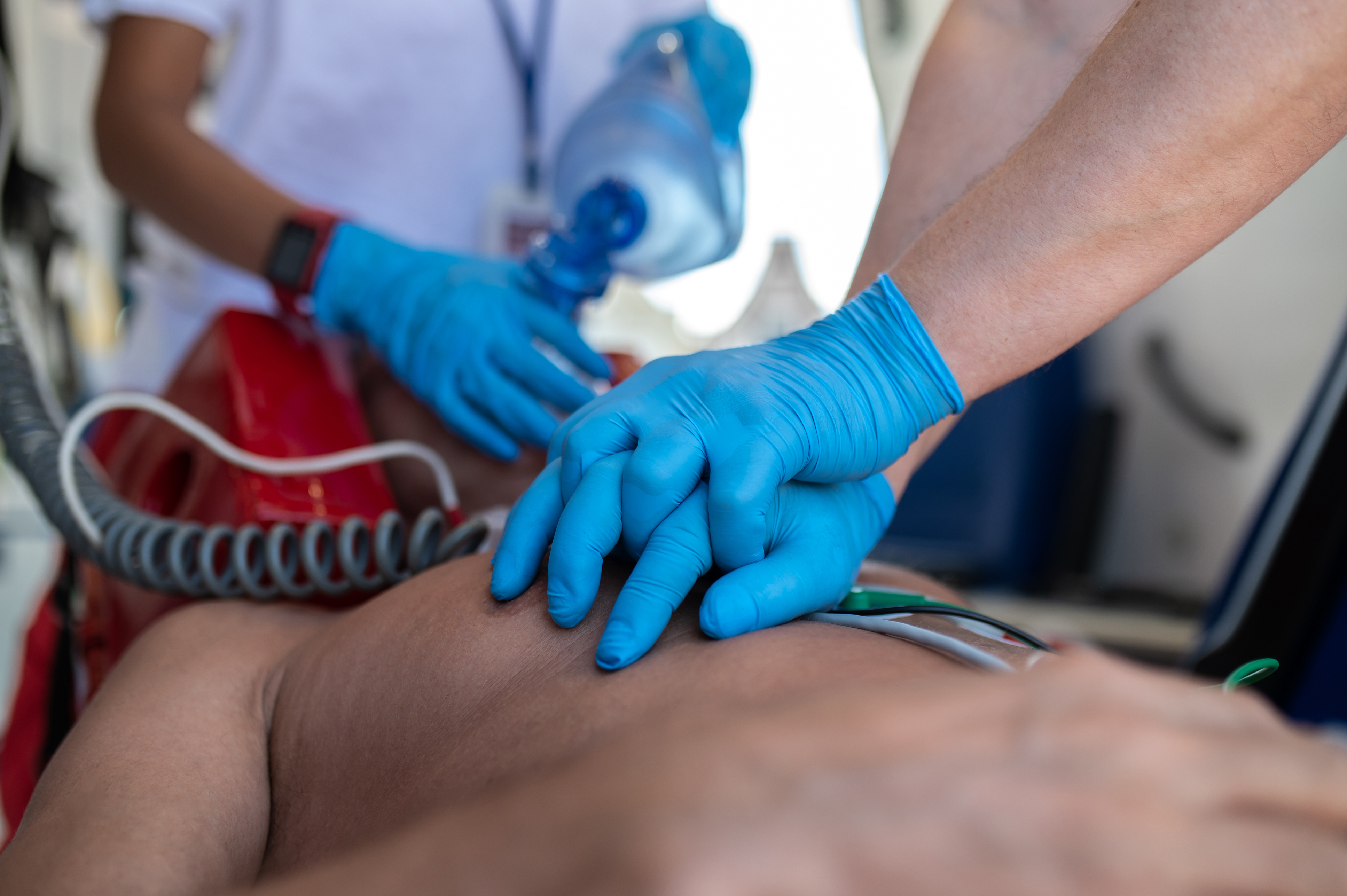 When a patient is in cardiac arrest, it means the heart has stopped functioning, thereby ceasing to provide oxygenated blood to all tissues. In this situation, nursing professionals must act swiftly.
Signs and symptoms of cardiac arrest include chest pain radiating to the left arm or back, diaphoresis, tachycardia, tachypnea, fatigue, dizziness, nausea, heart palpitations, or sudden loss of consciousness.
The American Heart Association (n.d.) has developed algorithms and provided free access to enable potential rescuers to learn and save lives. The algorithm, the link to which you will find in the recommended readings, is explained below.
For CPR the sequence is CABDE: Circulation, Airway, Breathing, Neurological Deficit, and Exam and Exposition.
In Circulation, the goal is to provide compressions at a rate of 100 to 120 bpm with a chest depth of 5 cm. The cycle starts with 15 compressions and two insufflations. After completing 4 cycles, pause to check the patient's pulse and ventilation. Remember that the cycles start with compressions before ventilation. If a VT/FT trace is present, administer defibrillation first, then compressions: 120 to 200 volts for biphasic and 360 volts for monophasic defibrillators.
IV lines are crucial for administering medications. If possible, install two lines with 14-16 gauge catheters. The drugs used in the protocol are epinephrine and amiodarone. Administer epinephrine at 1 mg every 3 to 5 minutes in cases of tachyarrhythmias. For amiodarone IV/IO, use a first dose of 300 mg bolus followed by a second dose of 150 mg. Lidocaine IV/IO can be administered at a first dose of 1-1.5 mg/kg and a second dose of 0.5-0.75 mg/kg.
You can ascertain that the patient has returned to spontaneous breathing if there is a pulse and blood pressure, an abrupt and sustained increase in PetCO2 (typically ≥40 mm Hg), or spontaneous arterial pressure waves observed with intra-arterial monitoring.
For Airway management, place the patient in a supine position on a hard surface and align the airway, ensuring there are no objects obstructing it.
For Breathing, provide two ventilations after 15 compressions. The Valve Bag Mask (VBM) is used for initial management, but Endotracheal Intubation (ET) or Supraglottic Advanced Airway should ultimately be utilized. Use waveform capnography or capnometry to confirm and monitor ET tube placement. Once the advanced airway is in place, administer one breath every 6 seconds (10 breaths/min), along with continuous chest compressions in intubated patients.
To assess Neurological Deficit, the Glasgow Coma Scale is useful to evaluate neurological status on recovering patients.
Finally, for Exam and Exposition, try to identify and reverse the cause. The most common causes are known as the 5 H’s and the 5 T’s: Hypovolemia, Hypoxia, Hydrogen ion (acidosis), Hypo-/hyperkalemia, Hypothermia, Tension pneumothorax, Tamponade - cardiac, Toxins, Thrombosis (pulmonary embolus), and Thrombosis (myocardial infarction).
4.3 Attending Acute Ischemic Stroke
 An ischemic stroke occurs due to the obstruction of tissue perfusion. In ischemia, blood flow is interrupted by a clot or an atheromatous plaque. In hemorrhage, perfusion is hindered by a lack of blood flow (American CPR, 2020).
An acute ischemic attack is one of the most common causes of death. The ischemic event, often due to a thrombus or an atheromatous plaque, obstructs blood flow. A hemorrhagic event involves the rupture of a cerebral artery, interrupting tissue perfusion.
Risk factors include high blood pressure, diabetes, smoking, dyslipidemia, and being over 60 years of age. Atrial fibrillation is a common and high-risk condition for a second ischemic stroke.
Both events are characterized by sudden neurological changes, including alterations in the state of consciousness, hemiparesis, loss of body strength, and facial asymmetry. Treatment depends on whether it was an ischemic or hemorrhagic event and on individual patient factors. In both cases, hemodynamic treatment following the ABCDE sequence is needed.
For Airway management, ensure proper positioning of the airway and maintain its permeability.
For Breathing, maintain an oxygen saturation (SaO2) level between 94-95%.
To manage Circulation, place the patient in the Fowler position, maintain blood pressure (BP) below 185/110 if thrombolytics are required, sustain normothermia, keep the glycemic index between 140 and 180 mg/dL, establish IV access, conduct lab tests, and continuously monitor blood glucose levels (American Heart Association, n.d.). Thrombolysis is indicated for ischemic events, depending on the patient's characteristics. The primary treatment for an ischemic stroke or cerebral infarction is IV Tissue Plasminogen Activator (tPA) if administered within the first 3 to 4.5 hours after the event. Surgery, including angioplasty and stent placement, may be necessary to dilate the artery and free the vessel's lumen (Kleindorfer et al., 2021).
In Neurological Deficit, there are two scales for pre-hospital assessment: the Cincinnati Pre-hospital Severe Stroke Severity Scale (CPSSS) and the National Institute of Health Stroke Scale (NIHSS). The CPSSS assesses facial palsy, asymmetric arm weakness, and speech disturbances, and each item can be scored as normal or not. If any of the three aspects is abnormal, the patient is suspected of having a stroke. The NIHSS assesses consciousness, eye movements, visual fields, motor and sensory impairments, ataxia, speech, cognition, and inattention (De Luca et al., 2019).
The Exam and exposition procedures allow us to identify possible alternative lesions suffered that need to be attended to.
Principal nursing care after treatment is composed of 4 main elements. These are the monitorization of the ECG traces to identify atrial fibrillation, monitorization of the BP to identify hypertension, promoting the Mediterranean diet (based on fruits, vegetables and less protein with low carbohydrates and sugar), and reinserting physical activity.
4.4 Managing the Care of a Client with Shock, Sepsis, and Trauma
 Shock is defined as "a state of organ hypoperfusion with resultant cellular dysfunction and death" (Procter, 2022).
This pathological entity is a state marked by tissue hypoperfusion resulting in tissue damage due to cell death and subsequent multiple organ dysfunction syndrome (MODS), which entails the failure of more than two organs and death. Principally, the lungs develop acute respiratory distress syndrome (ARDS), kidneys develop acute tubular necrosis, and the heart develops pump failure and disseminated intravascular coagulopathy (condition of the rapid proliferation of clots due to the low cardiac output).
The types of shock include hypovolemic, cardiogenic, distributive, and septic, each with a distinct cause.
Hypovolemic shock is a state of low corporal fluids due to bleeding or dehydration and is common in children.
Cardiogenic shock involves pump failure due to acute myocardial infarction and can also result from hindrances to adequate cardiac filling, such as in pericardial tamponade, pneumothorax, or valvar stenosis.
Distributive shock presents an excessive arteriolar vasodilation due to sepsis or systemic inflammatory response, such as anaphylaxis.
Finally, septic shock is understood as hypoperfusion due to alterations that are secondary to the arteriolar vasodilation caused by microorganisms. (Kislitsina et al., 2019).
Physical trauma is the most common cause of hypovolemic, cardiogenic, and distributive shock. Car accidents are regular causes of those traumas and figure among the first places on the list of morbidity worldwide.
In the early stages of shock, the skin becomes pale, mottled, and cyanotic. Then, the body tries to respond with compensatory mechanisms originated by the endogenous catecholamines such as epinephrine, norepinephrine, and dopamine. On subsequent stages of shock, the vital signs will drop down until failure.
Specific criteria for shock include obtundation, heart rate (HR) > 100 beats/minute, respiratory rate (RR) > 22 breaths/minute, systolic blood pressure (BP) < 90 mmHg or a 30 mmHg fall from baseline, and urine output < 0.5 mL/kg/hour. Laboratory findings that support the diagnosis include Lactate > 3 mmol/L (27 mg/dL), Base deficit < −4 mEq/L, and PaCO2 < 32 mm Hg (< 4.26 kPa) (Procter, 2022).
The following interventions must be both rapid and precise, as the patient's life depends on them. The ABCDE approach is once again utilized.
For Airway management, ensure the patient's mouth is free of obstacles such as teeth or foreign objects, bearing in mind that inflammation can obstruct the airway.
For Breathing, CO2 counteracts with the administration of high levels of oxygen. Considering the patient's severe health condition and potential airway obstruction from inflammation, rapid intubation is highly recommended for airway management. The team must know that and be prepared for it.
When managing Circulation, remember that peripheral vasoconstriction is a compensatory mechanism for hypovolemia or hypotension. Therefore, securing peripheral or central venous lines, and even intraosseous access, is crucial. Fluids should be replaced in case of hypovolemia and blood derivatives in case of bleeding. Corporal temperature must be conserved because patients in shock are cold because of the low circulation volume.
Common drugs used in shock include inotropes such as dobutamine or milrinone, vasopressors like vasopressin and epinephrine, stronger vasopressors such as norepinephrine, and amines like dobutamine or dopamine. When dealing with septic shock, antibiotics must be required usually in combination of ampicillin, polymyxin and cephalothin.
IV fluids used can include Ringer's lactate (which is isotonic), colloids, packed red cells, plasma, or plasma expanders. The goal is to maintain blood pressure above 90 mm Hg (Kaplan Nursing, 2023).
In assessing neurological deficits, the Glasgow Coma Scale is useful for identifying the patient's neurological impairment.
For Examination and Exposure, a detailed patient examination could identify the cause of shock, enabling its reversal. The 5 H’s and 5 T’s need to be specially monitored for identification of causes or consequences of shock and to be attended. ECG, vital signs and SaO2 must also be monitored. A urinary catheter should be placed to quantify renal urine output. Arterial gas exams, central venous pressure (CVP), serum electrolytes, blood urea nitrogen (BUN), creatinine, prothrombin time (PT), partial thromboplastin time (PTT), liver function tests, and fibrinogen and fibrin split products are done to monitor the patient’s physiological status and serve as a baseline for tracking (Procter, 2022).
Additional exams, such as X-rays, rapid bedside echocardiography, tomography, magnetic resonance imaging, or even infrared spectroscopy, may be required depending on the cause and type of shock. When dealing with septic shock, urinalysis, complete blood count, or cultures of blood, wound, or urine must be required. If the cause is not identified, a drug blood test or cardiac enzyme measurement will be performed to reach the closest assumed origin.
 In conclusion, patient care is a significant responsibility in all cases. Especially in emergencies, health personnel face the challenge of acting quickly and accurately to save lives. Having adequate action and a prompt response are two variables that can represent life or death for a patient. Studying important aspects such as the management of emergency situations in detail is crucial to fulfilling that responsibility.
- American CPR. (2020). ACLS Algorithms-updated. AHA Guidelines. https://americancprcertification.com/algorithms/acls-algorithms
- American Heart Association. (n.d.). Algorithms. https://cpr.heart.org/en/resuscitation-science/cpr-and-ecc-guidelines/algorithms
- De Luca, A., Mariani, M., Riccardi, M. T., & Damiani, G. (2019). The Role of the Cincinnati Prehospital Stroke Scale in the Emergency Department: Evidence from a Systematic Review and Meta-analysis. Open Access Emergency Medicine: OAEM, 11, 147-159. https://doi.org/10.2147/OAEM.S178544
- Kaplan Nursing. (2023). Physiological Integrity: Physiological Adaptation. NCLEX-RN Content Review Guide (9th ed.). Elservier. 39-46.
- Kislitsina, O. N., Rich, J. D., Wilcox, J. E., Pham, D. T., Churyla, A., Vorovich, E. B., Ghafourian, K., & Yancy, C. W. (2019). Shock: Classification and Pathophysiological Principles of Therapeutics. Current Cardiology Reviews, 15(2), 102-113. https://doi.org/10.2174/1573403X1566618121212502
- Kleindorfer, D. O., Towfighi, A., Chaturvedi, S., Cockroft, K. M., Gutierrez, J., Lombardi-Hill, D., Kamel, H., Kernan, W. N., Kittner, S. J., Leira, E. C., Lennon, O., Meschia, J. F., Nguyen, T. N., Pollak, P. M., Santangeli, P., Sharrief, A. Z., Smith, S. C., Turan, T. N., & Williams, L. S. (2021). 2021 Guideline for the Prevention of Stroke in Patients with Stroke and Transient Ischemic Attack: A Guideline from the American Heart Association/American Stroke Association. Stroke, 52(7), e364-e467. https://www.ahajournals.org/doi/full/10.1161/STR.0000000000000375#
- Procter, L. (2022). Shock. MSD Manual Consumer Version. https://www.msdmanuals.com/professional/critical-care-medicine/shock-and-fluid-resuscitation/shock
The following links do not belong to Tecmilenio University, when accessing to them, you must accept their terms and conditions.
Readings
- Mitchell, B. (2023). Overview of Arrhythmias. MSD Manual Consumer Version. https://www.msdmanuals.com/professional/cardiovascular-disorders/overview-of-arrhythmias-and-conduction-disorders/overview-of-arrhythmias
Videos
- RegisteredNurseRN.com. (2022, March 7). EKG/ECG Interpretation Basics Nursing NCLEX | QRS Complex, P Wave, T Wave, PR Interval [Video]. YouTube. https://www.youtube.com/watch?v=RoU4s18DXI4
La obra presentada es propiedad de ENSEÑANZA E INVESTIGACIÓN SUPERIOR A.C. (UNIVERSIDAD TECMILENIO), protegida por la Ley Federal de Derecho de Autor; la alteración o deformación de una obra, así como su reproducción, exhibición o ejecución pública sin el consentimiento de su autor y titular de los derechos correspondientes es constitutivo de un delito tipificado en la Ley Federal de Derechos de Autor, así como en las Leyes Internacionales de Derecho de Autor.
El uso de imágenes, fragmentos de videos, fragmentos de eventos culturales, programas y demás material que sea objeto de protección de los derechos de autor, es exclusivamente para fines educativos e informativos, y cualquier uso distinto como el lucro, reproducción, edición o modificación, será perseguido y sancionado por UNIVERSIDAD TECMILENIO.
Queda prohibido copiar, reproducir, distribuir, publicar, transmitir, difundir, o en cualquier modo explotar cualquier parte de esta obra sin la autorización previa por escrito de UNIVERSIDAD TECMILENIO. Sin embargo, usted podrá bajar material a su computadora personal para uso exclusivamente personal o educacional y no comercial limitado a una copia por página. No se podrá remover o alterar de la copia ninguna leyenda de Derechos de Autor o la que manifieste la autoría del material.